


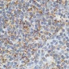
The Glysite™ Explorer in situ PLA Glycan Detection Kit is a fully integrated tool for the spatial detection of glycans proximal to a protein of interest in FFPE tissues, FFPE cell pellets and fixed cells. This system integrates a curated panel of Vector’s Glysite™ Explorer Lectins with Navinci’s proprietary in situ Proximity Ligation Assay (isPLA). Researchers can flexibly select specific Glysite™ Explorer Lectins covering the major categories of glycan types to pair with a primary antibody targeting their protein of interest. This provides a simple approach to understand glycan-protein proximity with a spatial context.
| Unit Size | 1 kit |
| Applications | Immunohistochemistry, Glycobiology |
| Sample Type | FFPE Tissue, FFPE Cell Pellet, Fixed Cells |
| Recommended Storage | Store reagents in original bottles at 4°C and -20°C accordingly |
| Unit Size | 0.1 mL per lectin |
| Applications | Immunohistochemistry, Glycobiology |
| Sample Type | FFPE Tissues, FFPE Cell Pellets, Fixed Cells |
| Recommended Storage | Store reagents in original bottles at 4°C. Do not freeze. |
| Recommended Usage | Each Glysite Explorer Lectin is sufficient to perform 50 reactions when using 100uL per reaction. Prior to use, Explorer Lectin (50x) should be diluted to 1x with Protein Diluent. |
| Sugar Specificity |
|
Glysite™ Explorer in situ PLA Glycan Detection Kit (GEK-1000) contains the following reagents:
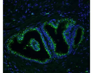
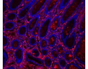
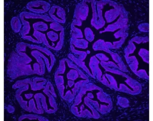
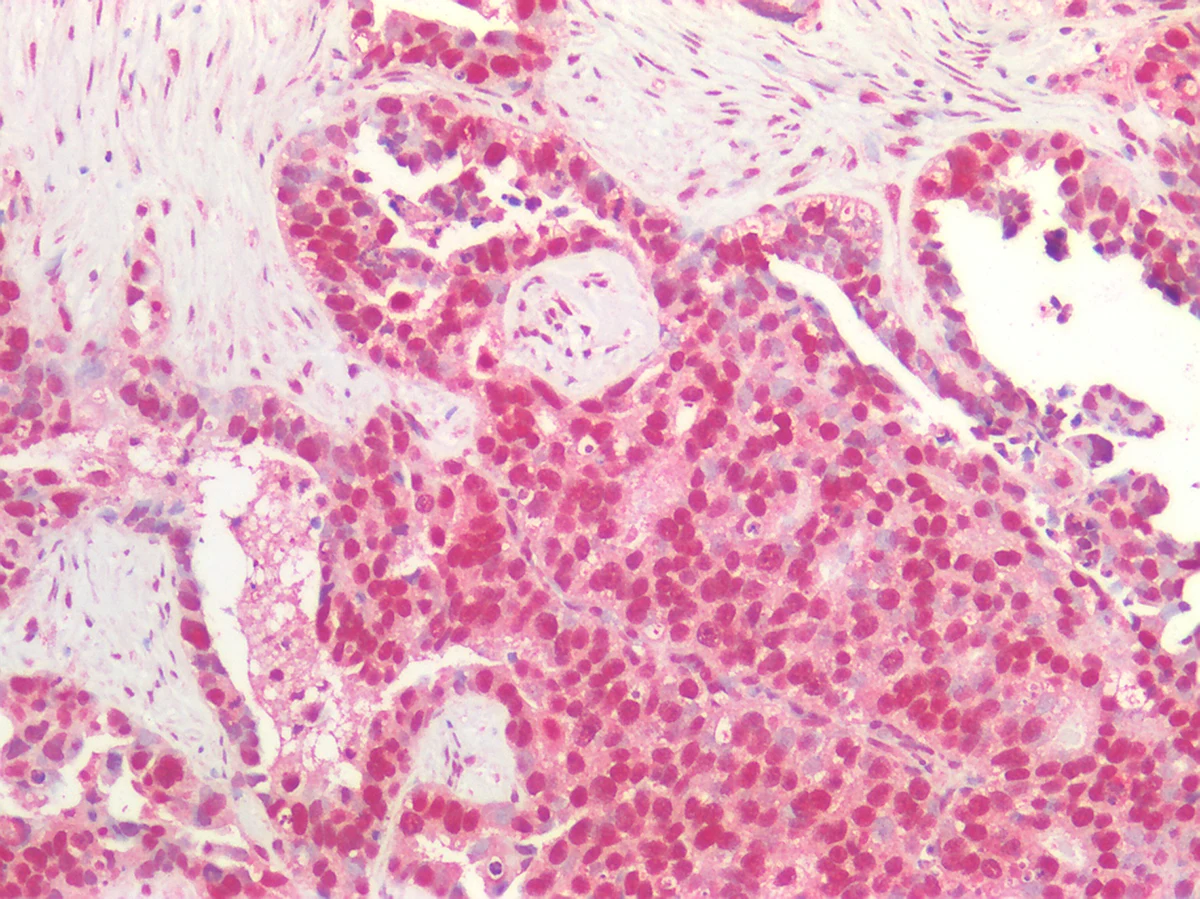
The Glysite Explorer Kit allows the visualization of a glycan and a protein when they are within 40nm of each other.
Customers will need to purchase a Glysite Explorer Lectin, and for additional reagent requirements, please see the User Guide.
The Glysite Explorer Kit has been validated on formalin-fixed, paraffin-embedded (FFPE) tissues, FFPE cells, and fixed cells. The kit is not recommended for use on frozen tissue specimens at this time.
Reagent volumes should be determined by the researcher to align with the dimensions of their specimens. Volumes sufficient to cover the specimens should be applied, and samples should not be allowed to dry out during the incubation steps.
The number of specimens that can be stained will vary depending on specimen size. The Glysite Explorer Kit provides 5 mL of working solution. If using 50-100µL of reagent per slide, there is sufficient volume to stain 50-100 specimens.
Slides stained with the Glysite Explorer Kit can be mounted with either an aqueous or a non-aqueous mounting medium. For non-aqueous mounting, we recommend using VectaMount® PT Permanent Mounting Medium (H-5600-60) or VectaMount® Express Mounting Medium (H-5700-60). For aqueous mounting, we recommend using VectaMount® AQ Aqueous Mounting Medium (H-5501-60).
The average run-time will depend on the duration of the primary antibody incubation. If your primary antibody incubation is 30 minutes, the total run time is approximately 8 hours.
If the assay cannot be completed within a single day, the tissue sections can be stored overnight at 4°C in Protein Diluent after incubation of the Lectin Solution in step 13. Do not allow the sample to dry out.
The kit is supplied with ImmPACT DAB, Vector’s most sensitive peroxidase substrate. Other peroxidase substrates can be substituted, and the Glysite Explorer Kit has been validated with ImmPACT® NovaRed (Catalog Number: SK-4805), ImmPACT® VIP (SK-4605), and ImmPACT® SG (SK-4705). However, not all peroxidase substrates are equivalent in sensitivity and staining quality and should be optimized to achieve desired results.
No, the Glysite Explorer Kit is not currently available with a fluorescent readout.
The Glysite™ Explorer in situ PLA Glycan Detection Kit leverages Vector’s well-known immunohistochemistry portfolio and lectins with Navinci’s proven isPLA technology. This assay enables researchers to detect glycosylation proximal to specific proteins in a spatial context.
![]()
Simplified scheme of Glysite™ Explorer in situ PLA Glycan Detection Kit. A. Lectin binds to target glycan and primary antibody binds to target protein. B. Oligo probes bind to primary antibody and lectin. C. When oligo probes are in proximity (~40nm), ligation occurs. D. Amplification and detection produces a colored precipitate at the site of proximity.
Recommended experiments before using the Glysite™ Explorer in situ PLA Glycan Detection Kit:
Specific. Sensitive. Reproducible.
